VIROLOGY
УДК 616.988-092.18-06:616-018.1
Z. A. Karalyan
Evolution of НЕр-2 cells under the influence of a chronic
infection of Оral
Polio Vaccine
(Submitted by academician K.G. Karageuzyan 13/VI 2003)
Chronically infected cell cultures are a
long-term associations between a host cells and viruses, characterized by long
coexistence of a virus and cell. The preservation of the viability of infected
cells during persistence is caused by the interaction of various viral and
cellular factors. During chronic viral infection a virus or cell or both are
changed. The evolution occurs through selection of more resistant population of
cells or less cytopathogenic virus [1]. Poliovirus can persistently infect some
human cell lines in vitro. Arising in vitro conditions of a chronic virus
infection by picornaviruses, and in particular by the poliomyelitis virus, is
caused by the appearance of resistant cell clones [2]. In such cells a virus is
replicated in lower titers compared to control cell without causing destructive
changes. The point of interest is the study of proliferative potential changes
in cancer cell cultures and degree of cells differentiation under the influence
of chronic virus infection.
Such parameters as the ploidy, size and the
quantity of nuclei and nucleoli are a valuable diagnostic attributes of the
transformed cells proliferation speed. [3,4,5].
Materials and methods. Cells. In work we used the transformed continuous cell culture of a human larynx
cancer - НЕр-2. Cells were cultivated in Eagle medium with glutamine and 10%
bovine serum. A monolayer of the intact cells was used in 48 hours after the
passage. Cells were resowed in dose 1×10⁵ cell/ml. The cell lines were received
from laboratory U 322 INSERM "Retrovirus et maladies associeees" Marseilles. The
choice of НЕр-2 as a model is motivated by the facts from publications on
susceptibility of these cells to the Sabin poliovirus strains.
Virus. In work was used
the standard trivalent Oral Polio Vaccine (OPV) (Polio SabinTM [oral] Poliomyelitis vaccine, live attenuated SB
BIOLOGICALS Rixensart - BELGIUM).
Chronic virus infection was received by one time infection of 48-hour monolayers of culture НЕр-2
by OPV. The multiplicity of infection was 0,000001 TCD50 per cell.
Infected cells were incubated at 36,5-370C. Persistent infection was
repeated 3 times and summarized data were presented. Viral titer was calculated
by the method of Karber. As a control the parallel conducted passages of
noninfected НЕр-2 culture were used. Their summarized data are given in the
table.
Quantitation of nuclear DNA. For the analysis of the received data, the cells preparations of НЕр-2
culture were fixed in 960 ethyl alcohol for 30 minutes and stained in
fresh Shiffs reactive, by Feulgen. The content of DNA in a nucleus and nucleolus
was defined by means of computer-equipped microscope-photometer SMP 05 (OPTON).
The television method was used on 575 nm waves. In each case 50 - 100 cells were
measured.
The DNA content is expressed in a "c" scale
in which 1c is half (haploid) the mean nuclear DNA content of cells from a
normal (non-pathological) diploid population in G0/G1 cell
cycle phase. The DNA image cytometric measurements identified cell nucleuses as
aneuploid if they deviate more than 10% from the 2c, 4c, 8c, 16 c, i.e. if they
are outside 2c±0,2, 4c±0,4,
8c±0,8, 16c±1,6. The number of
all cells in euploid regions of the DNA histogram rescaled by the mean
corrective factor of the tissue type under investigation (1.8c - 2.2c; 3.6c -
4.4c; 7.2c - 8.8c; 14.4-17.6) also was calculated [6].
Statistical analysis. For comparison of two groups the non-parametric U test analysis according
to Mann-Whitney was applied. Also statistical analyses were performed with
Student's t test in the SPSS version 8.0-computer software package (SPSS, Inc.,
Chicago, IL).
Results and discussion. After the infection, during 2-3 passages of OPV in cell culture,
cytopathogenic action of a virus was found as the degradation of the monolayer.
However, destruction of a monolayer was not complete, and after the 3rd passage
the cytopathogenic action of the virus was reduced. Received chronically
infected culture was characterized by insular and slower growth (approximately
twice), lack of capacity to form monolayer and constant allocation of a virus.
During 2nd 3rd passages, cytopathogenic action of a virus was found. After the
3rd passage the reduction of cytopathogenic action of a virus was observed. The
virus was found from the 1, 2, 3, 5, 9 passages in cell-free medium in titers
1,5-0,5 lg/ml. The highest titer was observed only once at 3-th passage (1,75).
During the further passages the virus was found out only in cells after their
destruction by freezing in low titres (0,25-0,75). A virus accumulated on
sensitive intact cell culture НЕр-2. So, low viral titres may be explained as a
result of decreased quantity of the sensitive cells in infected culture, and in
first passages as results of low multiplicity of infection. Quantity of the
cells in all passages after 2 was significantly less in comparison with control.
So in the 1-st passage, we can observe the
significant growth in the number of cells with 4 nucleoli, as well as the
tendency to increase in the percentage of cells with 5 nucleoli (0,72±0,08). This happened first of all due to significant
decrease in the number of cells with 1 nucleolus. From the 2-nd passage the
number of 4 nucleolar cells decreases and at the same time the quantity of cells
with 1 nucleolus increases. These processes continue and by the 5-th passage the
number of cells with 4 nucleoli sharply decreases, cells with 5 nucleoli almost
disappear (in following passages they percent vary from 0,07±0,01 up to 0,16±0,03). In
comparison with the control and the 1-st passage the quantity of cells with 1
nucleoli significantly raised. From 6-th passage the quantity of nonnucleolar
cells significantly increases, at practically the same parameters of cells of
other types. By the 12-th passage all these parameters almost do not changes.
Only the number of nonnucleolar cells in relation to the 6-th passage increases.
In this passage in relation to the control a significant decrease in the number
of 4 nucleolar cells and increase in the quantity of 1 nucleolar cells is
observed. In all the passages significantly increased the number of dead cells,
and by 12-th passage the quantity of mitoses significantly decreased. From the
6-th passage appeared significant number in comparison with the control of
nonnucleolar cells, the number of which continued to increase, and by the 12-th
passage it was significant not only in relation to the control, but also to the
6-th passage. At the same time the number of dead cells significantly increased
and the quantity of mitoses decreases (except the 5-th passage). Significant
changes in the quantity 2- and 3- nucleolar cells were not fixed in any passage
in comparison with the control.
Table 1
Nuclear DNA quantity in HEp-2 cells
under the influence of chronic viral infection
| Passage |
control |
1 |
5 |
6 |
9 |
12 |
| quantity on DNA (in conventional units) |
178.88 ±12.39 |
179.31 ±19.92 |
165.46 ±18.15 |
164.61 ±22.51 |
134.72 ±19.44 |
137.12 ±17.25* |
*Significant in comparison with the control p < 0,05
The received data testified that: under the
influence of a chronic viral functional activity of the tumour cells in culture
is decreased and in the greater degree the characteristics of their
differentiation were changed. The results of the study testify that the
proliferative activity of НЕр-2 cells was decreased under the influence of a
chronic viral infection.
Changes in DNA-ploidy were directly related
to the differentiation stage and cell proliferation that is why the quantity of
DNA in interphase nucleuses of НЕр-2 cell line (table 1) was also investigated.
We showed, that by the 12-th passage, under the influence of OPV this DNA
parameter significant decrease in nuclei of cells of a line НЕр-2.
We also investigated the quantity of DNA in
cells of each passage with various quantities of nucleoli. In our work it was
not revealed significant changes in the quantity DNA in cells with various
quantity of nucleoli. Thus, fluctuations of quantity of the nucleolar organizers
were not connected with the change of quantity of the DNA in the nucleus.
It was also investigated the area of
nucleoli, and difference in nucleoli between 1- 2 -, 3 -, 4 -, 5- nucleolar
cells and DNA quantity in nucleoli. In works [7] were showed that area of
nucleoli have a strong correlation with the changes of the cellular population
doubling time, so and with proliferation activity. It is known, that the
correlation of the number both size nucleolar organizer regions and quantity,
size and form of the nucleolus was determined by quantity, or by the
transcriptional activity of ribosomal DNA.
Table 2
Changes of nucleolar DNA quantity and
nucleolus square in HEp-2 cells
under the influence of chronic viral infection
| Pas- |
1 nucleolus |
2 nucleolus |
3 nucleolus |
4 nucleolus |
| sage |
DNA quantity |
square |
DNA quantity |
square |
DNA quantity |
square |
DNA quantity |
square |
| 1 |
19.9±2.7* |
7±1 |
23.4±4.9 |
9±2 |
27.4±7.1 |
10±3 |
35±2.7** |
14±4 |
| 5 |
20.3±4.3* |
7±2 |
24.4±3.6 |
9±2 |
29.7±4.7 |
11±2 |
26.1±4.2* |
11±1 |
| 6 |
21.8±3.8 |
8±2 |
24.9±3.9 |
9±2 |
33.4±7.3 |
12±3 |
32.1±4.1 |
11±3 |
| 9 |
21.5±4 |
8±2 |
22.2±6.1 |
8±2 |
26.3±4.1 |
10±2 |
27.5±5.5 |
10±2 |
| 12 |
22.7±3.3* |
8±2 |
26.1±5.9 |
9±2 |
30.2±8.1 |
11±3 |
32.5±4.1 |
12±1 |
| Cont |
28.8±3.9 |
10±3 |
27.1±5.1 |
10±3 |
29.4±6.1 |
11±2 |
37.7±2.3 |
13±2 |
*Significant in comparison with the control p <
0,05
**Significant in comparison with the 1 nucleolar cells p <
0,05
Our experiments have shown the reduction of
the DNA quantity in the nucleoli of mononucleolar cells in comparison with the
control after the beginning of influence of the chronic viral infection (tab.
2). This parameter is significant in the 1st, 5th, 7th and 12th passages. Other
passages show only a tendency to the decrease of the DNA quantity. The study of
4 nucleolar cells resulted in the fact that a significant decrease of the DNA
quantity in the nucleoli was observed only once in the 5th passages. Other
passages show only a tendency to the decrease of DNA quantity in the nucleoli.
Comparing mononucleolar and 4 nucleolar cells
of the same passage, we conclude that there is no significant difference in the
quantity of DNA of mononucleolar cells and total DNA of 4 nucleolar cells. There
were no significant changes in the 2 and 3 nucleolar cells either in comparison
with the control or inside each passage. The study of the nucleolar area of the
HEp-2 cells showed a tendency to the increase of the total nucleolar area in the
4 nucleolar cells.
All mentioned above testifies to the change
of nucleolar parameters of mononucleolar cells in the infected cells affected by
the chronic viral infection. This enables us to put forward the genetically
difference of these cells from the controlled ones.
The study of the nucleolar area revealed a
tendency to the increase of total nucleolar area in 4 nucleolar cells in
comparison with mononucleolar one, excluding the 1st passage where the increase
of the total DNA quantity is significant but in other cases only the tendency (t
varies from 0,88 to 1,94).
Distribution by the DNA ploidy and percentage
of the euploid cells also was studied. As noted in table 5 the increasing of the
number of euploid cells was observed at chronic infection. So, ploidy balance
(difference between the percentages of euploid and aneuploid cells) was
decreased. In table 3 shown the significant reduction of DNA ploidy during
chronic viral infection in HEp-2 cells which was observed since 9th passage.
Table 3
Changes in DNA ploidy in HEp-2 cells
during chronic viral infection
|
Passages |
|
control |
1 |
5 |
6 |
9 |
12 |
| % of the euploid cells |
12±3.1 |
20±3.9 |
18±5.2 |
18±4.5 |
34±7.1 |
34±5.3* |
| Average quantity of the DNA in "c" units |
5.96 |
5.98 |
5.52 |
5.49 |
4.51 |
4.57 |
*Significant in comparison with control and 1, 5, 6 passages
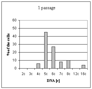
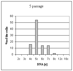
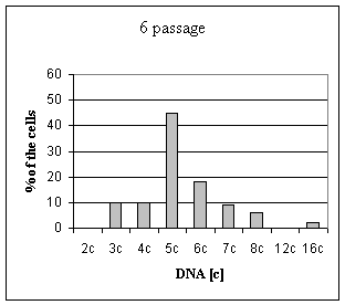
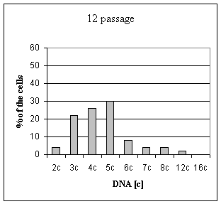
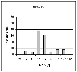
Fig 1 summarizes changes in DNA ploidy
indices during chronic viral infection. The present results indicate that under
the influence of OPV is decreased average ploidy of HEp-2 cells. The first time
the essential quantities of the diploid cells increases in 12th passage.
Percentage of aneuploid cells was decreased in 9 and 12 passages in comparison
with the control.
The present results indicate that there are
significant differences in various nuclear and nucleolar indices in HEp-2 cells
during chronic viral infection. Summing up, at 9 - 12 passages there is an
increase in the quantity of mononucleolar cells and accordingly decreases in
that of 4 nucleolar ones and stabilization in the quantity of 2 and 3 nucleolar
cells. There is also a significant decrease in the percentage of mitosis and
increase in the percentage of dead cells.
Figure 1. Distribution of the nucleus by the DNA ploidy
(in "c" units) in
HEp-2 cells during chronic viral infection (shown only a
significant
local maximum in the DNA histogram)
The evolution of cell population during
chronic viral infection in vitro condition takes place through selection of more
resistant population of cells or less cytopathogenic virus. Viability of
infected cells in vitro during persistence is caused by the interaction of
various viral and cellular factors. In our research the chronic viral infection
could be the result of action as cellular as viral factors because of in the
literature were presented data about unstable Sabin strains of poliomyelitis at
370C. However, the used virus keep citotoxic effect on sensitive
cells. In the other hand, in our experiment was shown significant difference
between intact cells and cells of 9-12 passages. Proliferative activity in this
population was sharply decreased. The chronically infected cells had a decreased
ploidy index and significantly increased number of the cells with euploid
quantity of the DNA in nucleus in comparison with control. These data allow to
assume, that under the influence of OPV there was a decrease of a proliferation
activity of HEp-2 cells and increase their differentiation [6].
The action of this factor was probably the
main reason of the change in nucleolar and nuclear parameters of the HEp-2
culture. It is known that the number of the nucleolar-forming regions in cells
is realized genotypically [8]. So, the changes in their quantity give us
possiblity to assume that the occurrence of more differentiated and less active
proliferating clones of cells is conditioned by the selective cytodestruction of
the less differentiated cells during the chronic viral infection. This
conclusion is based on the data that various mutations including virus-induces
are more dangerous for active divided cells, than for less active. This
supposition is made true by the significant changes of the nucleolar parameters
(the DNA quantity in nucleoli) and the increasing of the quantity of euploid
cells at comparing the control of the HEp-2 cell with the affected cells by
chronic infection of the OPV.
Institute of molecular biology NSA RA
Reference
1. Chiarini A., Arista S., Giammanco A., Sinatra A. - Journal of General Virology. 1983. V. 64.
P.1101-1110.
2. Kaplan G.,
Ricaniello V. - J. Virol. 1991. V. 65. N 4. P.
1829-1835.
3. Derenzini M., Trere
D., Pession A., Montanaro L., Sirri V., Ochs R. L. -
American Journal of Pathology. 1998. V. 152. P.
1291-1297.
4. Derenzini M., Trere
D., Pession A., Govoni M., Sirri V., Chieco P. - J Patholl.
1998. Jun. V. 191 (2). P. 181-6.
5. Friedrich K., Scheithauer J., Dimmer V., Meyer W., Theissig F., Haroske
G., Kunze K. D. - Anal Cell Pathol. 2000. V. 20 (2-3). P.
69-82.
6. Haroske G., Dimmer V.,
Meyer W., Kunze K.D. - Anal Cell Pathol. 1997. V. 15. P.
157-174.
7. Canet V., Montmasson
M.P., Usson Y., Giroud F., Brugal G. - Cytometry. 2001. Feb.
N 1. V.43(2). P.110-6.
8. Taylor
E. F., Martin-DeLeon P.A. - Amer. J. Hum. Genet. 1981. V.
33. P. 67-76.